May 19 2017
A Los Alamos research team have used the neutron crystallography technique to map the three-dimensional structure of a protein that breaks down polysaccharides, such as the fibrous cellulose of woody plants and grasses.
This finding could help cut the cost of producing biofuels. The research focused on lytic polysaccharide monooxygenases (LPMOs), a class of copper-dependent enzymes used by bacteria and fungi to naturally break apart cellulose and closely linked chitin biopolymers.
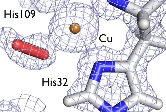
 Understanding the structure of an enzyme that helps bacteria break down cellulose and chitin in woody plant fibers can aid in developing better biofuels. In this image, an electron density map (gray) shows the structure of the active site center of the LPMO enzyme under study, depicting a dioxygen molecule (red stick) bound to a catalytic copper ion (bronze). Credit: Los Alamos National Laboratory
In the long term, understanding the mechanism of this class of proteins can lead to enzymes with improved characteristics that make production of ethanol increasingly economically feasible.
Julian Chen, Scientist, Los Alamos National Laboratory
A multi-institution team used the Advanced Light Source (ALS) synchrotron X-ray source at Lawrence Berkeley National Laboratory and the neutron scattering facility at the Spallation Neutron Source (SNS) at Oak Ridge National Laboratory to study LPMO. Both ALS and SNS are DOE Office of Science User Facilities.
Los Alamos Bioscience Division Researchers Chen, Clifford Unkefer, and former Postdoctoral Fellow John Bacik worked along with other Researchers from Lawrence Berkeley Laboratory, Oak Ridge National Laboratory, and the Norwegian University of Life Sciences to solve the structure of a chitin-degrading LPMO from the bacterium Jonesia denitrificans (JdLPMO10A). The study results are published in the Biochemistry journal (http://pubs.acs.org/doi/abs/10.1021/acs.biochem.7b00019?journalCode=bichaw).
The biggest challenge facing Biofuel Scientists is to identify cost-effective methods to break down polysaccharides, such as starches and cellulose that are widely spread in plants, into their subcomponent sugars for producing biofuel. LPMO enzymes, which are considered as key to this process, utilize a single copper ion to activate oxygen, a crucial step for the catalytic degrading action of the enzyme.
While the specific mechanism of LPMO action is unclear, it is assumed that catalysis involves initial formation of a superoxide through electron transfer from the reduced copper ion. By knowing the location of the copper ion and the constellation of atoms near it, the Scientists hope to reveal more about the enzyme’s function.
Although several X-ray crystallographic structures are presently offered for LPMOs from bacterial and fungal species, this new structure is more complete. The Researchers used X-ray crystallography in order to resolve the three-dimensional structure in detail of all the atoms except for hydrogens, the tiniest and most abundant atoms in proteins. The positions of hydrogen atom are vital for revealing functional characteristics of the target protein, and they can be visualized better using a neutron crystallography. The Researchers used this complementary method to determine the three-dimensional structure of the LPMO and to highlight the hydrogen atoms.
Notably, the crystallized LPMO enzyme has been caught while binding oxygen. In addition to the recent structures of LPMOs from different bacterial and fungal species, the results of this study points out a common mechanism of degrading cellulosic biomass even with differences in their protein sequences. This study has provided more insight into the mechanism of action of LPMOs, specifically the role of the copper ion and the nature of oxygen involvement.
This biofuels research is part of the Los Alamos National Laboratory’s mission that focuses on combining Research and Development solutions to attain the maximum impact on strategic National Security priorities, for example, New Energy sources.